Tibial fractures are a common occurrence, often caused by traumatic accidents or excessive force applied to the shinbone. It is the larger of the two leg bones. These fractures can vary in severity and location, each requiring specific diagnosis and treatment approaches. In this article, we’ll delve into the types of tibial fractures, tibial fracture nail, and orthopedic tools for effective treatment.
Anatomy of a Tibial Fracture
The anatomy of a tibial fracture involves the intricate structure of the tibia, or shinbone, when it experiences a break. The tibia is one of the two long bones in the lower leg, the other being the fibula. It plays a vital role in supporting body weight and assisting in various movements. Tibial fracture affects the bone’s integrity. It can also impact adjacent structures such as muscles, blood vessels, and nerves.
The tibia is divided into three main sections:
Proximal Tibia: This is the upper part of the tibia, which connects to the knee joint. It includes the tibial plateau, a flat area that forms the top surface of the bone, and the tibial tuberosity, a bony prominence just below the knee joint to which the patellar tendon attaches.
Shaft of the Tibia: The shaft is the long, middle portion of the tibia. It is the largest and most weight-bearing part of the bone. Fractures in this region can vary in nature, such as transverse, oblique, or spiral fractures, depending on the direction of the break.
Distal Tibia: This is the lower part of the tibia, which connects to the ankle joint. It forms the prominence of the inner ankle and plays a significant role in stabilizing the ankle.
Classification of Tibial Fracture
When a tibial fracture occurs, it can be classified based on various factors:
Open vs. Closed: An open fracture involves a break in the skin, allowing the bone to be exposed to the external environment. A closed fracture, on the other hand, does not break the skin.
Complete vs. Incomplete: A complete fracture involves a break that extends through the entire width of the bone, resulting in two separate fragments. An incomplete fracture, also known as a greenstick fracture, involves a crack in the bone without complete separation.
Displaced vs. Non-displaced: A displaced fracture involves the bone fragments being out of their normal alignment, while a non-displaced fracture means the bone pieces are still relatively aligned.
Comminuted: A comminuted fracture is characterized by the bone being shattered into multiple pieces.
Types of Tibial Fractures
Tibial fractures can be broadly categorized into three main types:
Fractures of the Proximal Tibia: These fractures occur near the knee joint and are typically classified as either fractures of the tibial plateau (the flat top of the shinbone) or fractures of the tibial tuberosity (the bony bump below the knee cap).
Fractures of the Shaft of the Tibia: These fractures occur along the long, middle portion of the shinbone. They can be further subdivided into transverse fractures (straight across the bone), oblique fractures (diagonal break), and spiral fractures (twisting break).
Fractures of the Distal Tibia: These fractures occur closer to the ankle joint and are often more complex due to the proximity of the ankle joint and the intricate network of ligaments and tendons in the area.
Diagnosis of Tibial Fractures
Diagnosing a tibial fracture involves a combination of physical examination, medical imaging, and patient history assessment. Doctors will evaluate the patient’s symptoms, conduct a thorough physical examination, and order appropriate imaging tests such as X-rays, CT scans, or MRI scans. These imaging techniques allow physicians to assess the location, severity, and complexity of the fracture, which is crucial for determining the most suitable course of treatment.
Tibial Fracture Tools for Treatment
The treatment of tibial fractures often involves surgical intervention, during which orthopedic implants play a vital role in stabilizing the broken bone and promoting proper healing. Commonly used implants include:
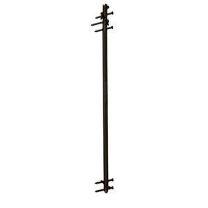
- Intramedullary Nails (IM Nails): Tibial nails, also known as intramedullary nails are typically made of biocompatible materials such as stainless steel or titanium. Medical professionals insert these long, slender rods into the marrow canal of the tibia, the larger of the two bones in the lower leg. This method, known as “Intramedullary nailing,” involves placing tibial nails within the bone’s medullary canal.

- Locking Plates: Orthopedic plates are metal implants designed to provide stability to fractured bones by holding the broken segments in their correct positions. They come in various shapes and sizes to accommodate different bone structures and fracture patterns. Typically, manufacturers make plates from materials such as stainless steel or titanium. These plates are
biocompatible. They provide strength while minimizing the risk of allergic reactions.
- Locking Screws: Locking screws are specialized screws used in conjunction with orthopedic plates, especially in cases where stable fixation is crucial. Unlike traditional screws, which compress the bone fragments against the plate, locking screws create a fixed-angle construct. This construct provides more resistance to movement. It is particularly useful in cases of poor bone quality or when dealing with complex fractures.
- External Fixators: In cases of severe open fractures or fractures with extensive soft tissue damage, external fixators may be used. Placing these devices outside the body and connecting them to the bone fragments via pins creates a stable external frame.


Benefits of Tibial Nails
Tibial nails offer several advantages in the treatment of fractures:
- Stability: The nail’s placement within the bone provides strong support and stability to the fractured segments, promoting optimal alignment and healing.
- Load Sharing: Tibial nails allow for load sharing between the implant and the bone, enabling early weight-bearing and ambulation, which can contribute to faster recovery.
- Minimized Soft Tissue Disruption: Inserting the nail within the medullary canal reduces the need for extensive soft tissue dissection, resulting in less damage to surrounding structures and potentially faster healing.
- Preservation of Blood Supply: The implantation of tibial nails does not disrupt the bone’s blood supply, which can aid in the healing process.
Benefits of Locking Plates
- Enhanced Stability: One of the primary advantages of locking plates is their ability to provide enhanced stability to fractured bones.
- Versatility: Locking plates are versatile, ideal for diverse orthopedic uses, and effective for fractures, even complex comminuted ones, lacking traditional screw stability.
- Improved Fracture Healing: The stability provided by locking plates contributes to improved fracture healing.
- Reduced Risk of Screw Cut-Out: Traditional plates use screws that compress the bone against the plate, which can sometimes lead to the phenomenon known as “screw cut-out.”
- Benefit in Poor Bone Quality: Locking plates are especially advantageous in cases of poor bone quality, such as osteoporosis.
Conclusion
Zealmax Ortho shines as a dedicated manufacturer and exporter of orthopedic tibial fracture tools. Their expertise is particularly evident in the realm of tibia fracture tools. Zealmax Ortho plays a pivotal role in enhancing patient outcomes. We are contributing to the evolution of orthopedic practices on a global scale. We are offering advanced solutions tailored to the intricate needs of tibia fracture treatment.
