The femur is the human body’s longest, strongest, and heaviest tubular bone, as well as one of the primary load bearing bones in the lower extremity. High energy forces, such as car accidents, frequently cause femoral shaft fractures. Adult midshaft femur fracture complications and injuries can be fatal, including hemorrhage, internal organ injury, wound infection, fat embolism, and adult respiratory distress syndrome.
Femoral shaft fractures can also cause significant physical impairment due to the possibility of fracture shortening, malalignment, or prolonged immobilization of the extremity with casting or traction. The art of femoral fracture care entails a delicate balancing act between anatomic alignment and early functional limb rehabilitation.
Causes of Femoral Shaft Fractures
Femoral shaft fractures in children are frequently caused by a high-energy collision. A car or motorcycle accident is the most common cause of femoral shaft fracture. Another common cause is being hit by a car while walking, as are falls from great heights and gunshot wounds.
In an older person with weaker bones, a lower-force incident, such as a fall from standing, may result in a femoral shaft fracture.
Most common risk factors
Midshaft femur fractures occur at a rate of about 10 per 100,000 person-years. The incidence rises in the young, then falls after the age of 20, and then rises again in the elderly. Over the age of 75, there is a significant increase. The majority of femur fractures occur in the proximal third (also known as hip fractures), which are discussed in detail elsewhere.
Young men are most likely to sustain femoral fractures from severe trauma, especially diaphyseal fractures. Patients under the age of 40 have a higher risk of suffering high energy trauma (such as a car accident) and fracturing the midshaft of the femur, whereas patients over 40 have a higher risk of suffering low energy trauma (such as a fall) and fracturing the proximal third of the femur. Eighty percent of people aged 35 or older who suffered femur fractures from moderate energy trauma had a history of generalized osteopenia or a condition that was likely to do so. Low-energy falls are the leading cause of fractures in older adults, accounting for 65% of all fractures.
These frequently happen at home. The risk of femur fracture may increase with long-term bisphosphonate use. Most other femur fractures result from gunshot wounds and industrial accidents.
Symptoms of Femoral Shaft Fractures
A femoral shaft fracture usually causes severe pain right away. You will be unable to bear weight on the injured leg, and it may appear deformed—shorter than the other leg and bent. Some of the symptoms of a femur shaft fracture include:
- Severe and sharp pain
- Inability to put weight on the injured leg
- Swelling
- Bruising
- Pain on touching the thigh that worsens with movement
- Deformity
- Numbness in the thigh, lower leg, ankle, foot, knee
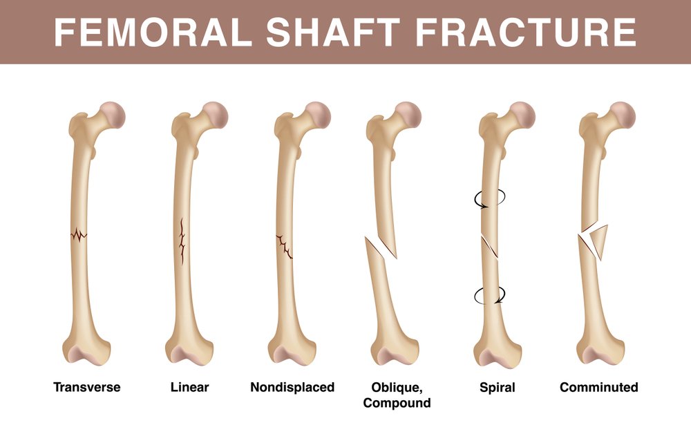
Types of Femoral Shaft Fractures
The force that causes the break in the femur determines the severity of the fracture. The bone fragments may line up correctly (stable fracture) or be misaligned (displaced fracture). The skin around the fracture may be intact (closed fracture) or punctured by the bone (open fracture). Femur fractures are classified depending on:
- The fracture’s location (the femoral shaft is divided into thirds: distal, middle, proximal).
- The fracture’s pattern (for example, the bone can break in different directions, such as crosswise, lengthwise, or in the middle).
- Whether the injury tore the skin and muscle over the bone.
The most common types of femoral shaft fractures include:
Transverse fracture
The break in this type of fracture is a straight horizontal line that runs across the femoral shaft.
Oblique fracture
An angled line runs across the shaft of this type of fracture.
Spiral fracture
Like the stripes on a candy cane, the fracture line encircles the shaft. This type of fracture is caused by a twisting force to the thigh.
Comminuted fracture
The bone has broken into three or more pieces in this type of fracture. The number of bone fragments usually corresponds to the amount of force required to break the bone.
Open fracture
An open or compound fracture occurs when a bone breaks in such a way that bone fragments protrude through the skin or a wound penetrates down to the broken bone. Open fractures frequently result in significantly more damage to the surrounding muscles, tendons, and ligaments. They are more prone to complications, particularly infections, and heal more slowly.
Diagnosis of Femoral Shaft Fractures
X-rays
X-rays are the most popular method of evaluating fractures because they give distinct images of the bone. X-rays can demonstrate whether a bone is broken or not. Additionally, they can display the kind of fracture and its location within the femur.
Computerized tomography (CT) scans
After examining your x-rays, if your doctor feels that more information is still required, he or she might request a CT scan. Your limb can be seen cross-sectionally during a CT scan. It may give your doctor important details about the fracture’s severity. For instance, the fracture lines can occasionally be very thin and challenging to see on an x-ray. Your doctor can see the lines more clearly with the aid of a CT scan.
Surgical Treatment of Femoral Shaft Fractures
Timing of surgery
The majority of femur fractures heal in 24 to 48 hours. Fixation may occasionally be postponed until other potentially fatal wounds or unstable medical conditions are stabilized. As soon as you get to the hospital, open fractures are given antibiotic treatment to lower the chance of infection. During surgery, the tissues, bone, and open wound will be cleaned.
Your doctor may decide to traction your leg or place it in a long-leg splint for the period of time before your surgery. This preserves the length of your leg and keeps your broken bones as aligned as possible. A pulley system of weights and counterweights called skeletal traction holds the fragments of broken bone together. It helps to keep your leg straight and frequently eases pain.
External fixation
Metal pins or screws are inserted into the bone above and below the fracture site during this type of procedure. The bar outside the skin is where the pins and screws are attached. The frame-like object holding the bones in place is a stabilizing device.
Femur fractures are typically treated temporarily with external fixation. External fixators are frequently applied when a patient has multiple injuries and is not yet prepared for a longer surgery to repair the fracture because they are simple to put on. Until the patient is fit enough for the final surgery, an external fixator offers good, temporary stability. An external fixator may occasionally be left in place until the femur has fully recovered, but this is uncommon.
Intramedullary nailing
Currently, intramedullary nailing is the procedure that most surgeons use to treat femoral shaft fractures. A specially crafted metal rod is inserted into the femur’s canal during this procedure. To hold the fracture in place, the rod crosses it.
Either the hip or the knee can be used to insert an intramedullary nail into the canal. To keep the leg in the right position while the bone heals, screws are inserted above and below the fracture. The most common material for intramedullary nails is titanium. They are available in a range of lengths and diameters to fit the majority of femur bones.
Plates and screws
The bone fragments are first moved (reduced) into their usual alignment during this procedure. Metal plates and screws that are affixed to the bone’s exterior hold them together. When intramedullary nailing may not be possible, such as for fractures that extend into the hip or knee joints, plates and screws are frequently used.
Conclusion
Zealmax Ortho is a trusted option for femur shaft fractures because it offers orthopedic implants that guarantee a quicker healing process, less pain, and a lower risk of complications. We are committed to the invention, design, and development of cutting-edge implants that restore function to lives and are novel, clinically applicable, and state-of-the-art.

