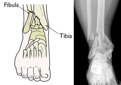
Pilon fractures are rare but severe bone breaks happen at the bottom of your shinbone near your ankle. They’re typically caused by high-impact events such as vehicle accidents. Treatment usually involves surgery.
Pilon fractures can range from a simple crack in the bone to a complex break with multiple fragments and significant displacement. They are often accompanied by soft tissue injuries, such as ligament damage or bruises to the surrounding muscles and tendons. In severe cases, Pilon fractures can also cause damage to the joint surfaces and surrounding nerves and blood vessels.
The most common cause of Pilon fractures is a high-energy trauma, such as a car accident or fall from a significant height. Pilon fractures are also more likely to occur in older individuals, as the bones become weaker and more brittle with age.
What is a pilon fracture?
A pilon fracture is a relatively uncommon bone break that occurs near your ankle at the bottom of your tibia (the larger of the two bones in your lower leg, or your shinbone). In many pilon fractures, the other bone in your lower leg, the fibula, is also broken. The majority of pilon fractures are caused by high-impact events, such as a car accident or a fall from a great height.

The word “pilon” in French refers to the pestle, an instrument with a rounded end used to crush and grind materials. Because of the crushing force that frequently results in them, this kind of bone break is known as a pilon fracture. Your talus, the bone that supports your weight in your ankle, is where your tibia and fibula are joined. Pilon fractures occur when the force of your talus striking your tibia causes your tibia (and frequently your fibula) to shatter at the ankle joint.
Different types of pilon fractures?
Depending on the shape of the break, pilon fractures can be classified using a variety of different categorization schemes. The following forms of pilon fractures are classified by healthcare professionals according to the Ruedi-Allgower classification system:
Type I: An articular fracture (bone break in or near a joint) is referred to as a pilon fracture if there is little to no bone displacement (the broken bones are still aligned).
Type II: A type II pilon fracture occurs when the articular surface of the bottom of the tibia is misaligned or displaced, but there are few or no pieces (communition). When the bone fractures into more than two pieces, it is known as a comminuted fracture.
Type III: When a bone has fractured into more than two pieces (comminution) and the ends of the broken bones have forced into one another, the fracture is classified as type III (impacted fracture). Type III fractures make up between 25% and 71% of pilon fractures.
There are also different types of fractures that can occur when a bone breaks. Your healthcare provider may refer to your pilon fracture using one or more of the following terms:
Closed or open (compound) fracture: A closed fracture occurs when the fracture does not break open the surrounding skin. An open fracture or a compound fracture occurs when a broken bone pierces the skin. Open fractures account for approximately 20% of pilon fractures.
Complete fracture: A complete fracture occurs when the bone is broken into two pieces.
Displaced fractures: A displaced fracture occurs when the broken bones do not remain aligned as they should.
Comminuted fracture: A comminuted fracture occurs when the bone breaks into more than two pieces.
Impacted fracture: An impacted fracture occurs when the ends of the broken bone are driven into each other.
Spiral fracture: A spiral fracture occurs when the fracture spirals around the bone.
Severity of pilon fractures
Pilon fractures differ. The tibia may break in a single piece or shatter into several pieces.
The severity of the injury is determined by a number of factors, including:
- The quantity of fractures.
- The number and size of broken bone fragments.
- The quantity of each piece is out of place (displaced). In some cases, the broken ends of bones almost perfectly line up; in more severe fractures, there may be a significant gap between the broken pieces, or the fragments may overlap.
- Injury to the soft tissues surrounding the injury, such as muscle, tendons, and skin.
The fracture is referred to as an “open” or complicated fracture if bone pieces protrude through the skin or if a wound extends down to the broken bone. Due to the possibility of infection in both the wound and the bone once the skin is broken, this sort of fracture is extremely dangerous. To stop infection, immediate therapy is necessary.
Cause of pilon fractures
Most frequently, high-energy trauma like a vehicle or motorcycle accident, a fall from a height, or a skiing accident causes pilon fractures.
Since air bags were first included in cars, doctors have noticed a rise in pilon fractures. More people can survive high-speed car crashes thanks to air bags, but because the legs are not protected, many of the survivors suffer from pilon fractures and other leg injuries.
Symptoms of pilon fractures
The symptoms of a Pilon fracture can include severe pain and swelling in the ankle, difficulty bearing weight on the affected foot, and bruising and tenderness in the area. In some cases, there may be deformity or misalignment of the ankle joint. Numbness, tingling, or weakness in the foot or lower leg may also be present if there is nerve or vascular damage.
If you suspect that you have a Pilon fracture, it is important to seek medical attention as soon as possible to receive a proper diagnosis and appropriate treatment. Early intervention can help to prevent long-term complications and promote a faster, more complete recovery.
Diagnosis of pilon fractures
Physical Examination
- Examine your lower leg and ankle, checking for wound cuts and carefully pressing various regions to feel for pain.
- Verify your ability to move your toes and feel various parts of your foot. In some circumstances, a bone break may also cause nerve damage.
- To ensure that your foot and ankle are receiving enough blood, take your pulse at several strategic locations on your foot.
Imaging Tests
X-rays– Images of dense things, like bone, are produced by X-rays. To assess a pilon fracture, it is normal practice to take X-rays of the leg, ankle, and foot. An x-ray can reveal whether your bones have been injured or whether the joints in your ankle are misaligned.
Computerized tomography (CT) scans– A CT scan can provide valuable information about the severity of the fracture by helping your doctor see the fracture lines more clearly. A CT scan will also help your doctor plan your treatment. Your doctor may order a CT scan right away, or may wait until later in your treatment — after an external fixator is applied.
Surgical treatment of pilon fractures
Surgery is commonly recommended for unstable fractures with out-of-place bones.
Open Reduction and Internal Fixation
During this procedure, the displaced bone fragments are repositioned (reduced) into their normal alignment before being held together with screws and metal plates attached to the bone’s outer surface.
External Fixation
Your doctor may use an external fixator to stabilize your ankle and hold your pilon fracture in place until your second surgery.
Conclusion
Zealmax Ortho was founded in 2006 with the exclusive goal of providing our consumers with world-class implants and instruments. Our objective is to develop health care technology and reduce health inequalities by delivering genuine, high-quality, and complete products while maintaining fairness, honesty, and corporate responsibility.

